Two Vessel Cord Radiology
A good alternative for PDW is STIR. The journals publications cover all imaging modalities radiology issues related to patients policy and practice improvements and clinically-oriented imaging physics and informatics.

Isolated Fetal Choroid Plexus Cysts Plexus Products Fetal Cysts
To Control Bleeding As an alternative to surgery interventional radiology may be used to control bleeding hemorrhage in conditions ranging from gastrointestinal bleeding to postpartum bleeding to trauma.

Two vessel cord radiology. Our students work with world-class faculty at top clinical and research centers affiliated with the University of Washington. Primary intra-axial tumors are usually derived from. Cerebral white matter diseases are classified into two broad categories. M1 Segment of the MCA. 3D Blood Vessel Map Reveals Location of Stem Cells. A radiographic technique in which a radiopaque shows up on X-ray contrast material is injected into a blood vessel for the purpose of identifying its anatomy on an X-rayThis technique is used to image arteries in the brain heart kidneys gastrointestinal tract aorta neck carotids chest limbs and pulmonary circuit.
Demyelination is an acquired disorder that affects normal myelin. A head MRI is an excellent way to diagnose whether a stroke is ischemic or hemorrhagic and its also great at finding abnormalities in the skull and spinal cord. The journal is a valuable resource for practicing radiologists radiologists-in-training and other clinicians with an interest in imaging. Make sure to have two different sequences or two different planes. Use a 512 matrix and cardiac gating for optimal results. It may be used to control or prevent abnormal bleeding close off vessels supplying blood to a tumor eliminate abnormal connections between arteries and veins or to treat.
Anatomically the MCA is divided into two segments M1 and M23. Periventricular white matter changes. Small vessel ischemic disease. Embolism Definition An embolism is an obstruction in a blood vessel due to a blood clot or other foreign matter that gets stuck while traveling through the bloodstream. This International journal Journal of Clinical Neuroscience publishes articles on clinical neurosurgery and neurology and the related neurosciences such as neuro-pathology neuro-radiology neuro-ophthalmology and neuro-physiology. Spinal cord tumors are uncommon and approximately 23 of primary intra-axial tumors of the central nervous system occur in the spinal cord.
Soft X-Ray Tomography for Quick 3D Imaging of Cell Organelles. JVIR published continuously since 1990 is an international monthly peer-reviewed interventional radiology journal. While CT scans can only provide images from one orientation MRIs can produce multiple pictures in several orientations. On PDW-images the spinal cord has a uniformly low signal intensity like CSF which gives the MS lesions a good contrast against the surrounding CSF and normal cord tissue. Much of the bone pain caused by the ABC is usually relieved within one or two treatments. Baltzer serves as a professor in radiology at the Department of Biomedical Imaging and Image-guided therapy at the Medical University of Vienna Austria.
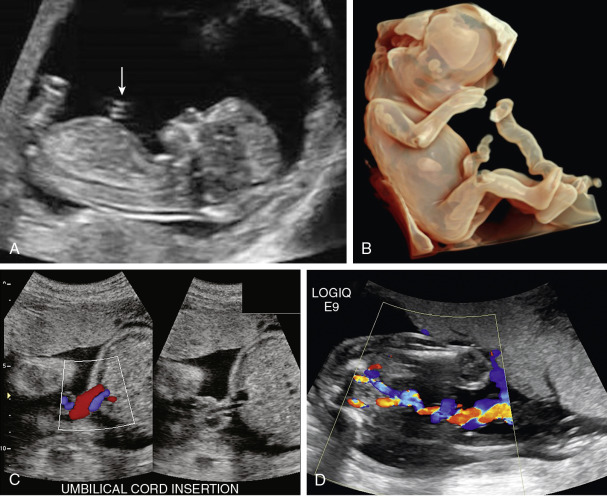
December 6th 2021. This is sometimes also called a two-vessel umbilical cord or two-vessel cordApproximately this affects between 1 in 100 and 1 in 500 pregnancies making it. New Hydrogel Material for Vocal Cord Repair. Via interventional radiology a radiologist may place a stent graft in the region of an aneurysm thus essentially relining the blood vessel. Schedule Your MRI or CT Scan Today. Each image used in this chapter was obtained using two-dimensional 2D ultrasound.
Risk stratification primary factors General schema for stratification Prognostic factors which dont add much Risk stratification lysis without a CT scan Physiology of the PE death spiral Resuscitation Avoid procedures if possible Fluid management Inotropes. As the official journal of the Society of Interventional Radiology JVIR is the peer-reviewed journal of choice for interventional radiologists radiologists cardiologists vascular surgeons neurosurgeons and other clinicians who seek current and reliable information on. UW Radiology is a top tier academic medical program with robust offerings for students residents and fellows. CONTENTS Rapid Reference Preamble Diagnosis risk stratification Is PE driving the patients instability. Now perhaps the best technical term for what Im referring to is cerebral small vessel disease But many other synonyms are used by the medical community especially in radiology reports. In a prospective multicenter study two blinded raters independently.
Michaels Hospital University of Toronto Canada Publicationdate 2008-07-02 This review is based on a presentation given by Walter Montanera and was adapted for the Radiology Assistant by Robin Smithuis. Materials and Methods This study was approved by the institutional review board. The discrepancy between the two becomes increasingly apparent in the mid-to-low regions of the thoracic spinal cord where a fracture at thoracic level 8 T8 might cause a neurological SCI at T12. Catheter embolization places medications or synthetic materials called embolic agents through a catheter into a blood vessel to block blood flow to an area of the body. Baltzer has ample editorial experience as he worked in various editor positions at the EJR European Radiology and PLoS One as well as guest editor for Der Radiologe. Spinal cord tumors are a challenge for patients and neurosurgeons because of the high risk of neurologic deficits from the disease process and surgical interventions.
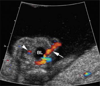
Occasionally there is only the one single umbilical artery SUA present in the umbilical cord. Allowing pulse wave assessment of this vessel for aneuploidy and cardiac screening. This segment is also called the horizontal segment or. The interventional radiology treatment of aneurysmal bone cyst involves a series one every 3 months of minimally invasive treatment injections of a drug called doxycycline. Learn more about spinal cord injury levels treatments rehabilitation symptoms causes diagnosis and how the injury will affect the rest of the body. All individuals provided signed informed consent.
Description Emboli have moved from the place where they were formed through the bloodstream to another part of the body where they obstruct an artery. The vast majority of white matter diseases especially in the adult fall into this category and are the principal focus of this chapter. Purpose To provide normal values of the cervical spinal canal and spinal cord dimensions in several planes with respect to spinal level age sex and body height. However in radiology and surgery the middle cerebral artery is divided into four parts M1 M2 M3 and M44. The plural of embolism is emboli. The therapy is performed as an outpatient procedure with minimal recovery time.
Interventional radiology IR is a medical subspecialty that performs various minimally-invasive procedures using medical imaging guidance such as x-ray fluoroscopy computed tomography magnetic resonance imaging or ultrasoundIR performs both diagnostic and therapeutic procedures through very small incisions or body orificesDiagnostic IR procedures are those. Radiology Department of the Rijnland hospital Leiderdorp the Netherlands and the Division of Neuroradiology of the St. Trimester and that deserves special mention is physiologic herniation of the midgut into the root of the abdominal cord insertion. The journal has a broad International perspective and emphasises the advances occurring in Asia the Pacific Rim. Spinal cord injury can cause a range of symptoms including weakness loss of muscle function and loss of sensation.

Posterior Fossa Radiology Epidermoid Cyst Neurology

Single Umbilical Artery Radiology Reference Article Radiopaedia Org

Single Umbilical Artery Radiology Reference Article Radiopaedia Org

The Fetal Three Vessel And Tracheal View Revisited Semantic Scholar Obstetric Ultrasound Cardiac Sonography Sonography Student

Shoulder Imaging Radiology Imaging Medical Radiology
Umbilical Cord Radiology Key

Omphalomesenteric Duct Cysts Sonography Ultrasound
Umbilical Cord Radiology Key
Umbilical Cord Radiology Key

Digital X Ray Centre In Ahmedabad In 2021 X Ray Radiation Dose Digital

Normal Fallopian Tube In Two Different Patients A Transvaginal Ultrasound Image Of An Elongated View Of A Nor Transvaginal Ultrasound Fallopian Tubes Ovaries

Magnetic Susceptibility Artifact Radiology Reference Article Radiopaedia Org Radiology Artifacts Brain Scan

Single Umbilical Artery Two Vessel Cord No Mickey Mouse In 2021 Mickey Mouse Batman Mickey
Single Umbilical Artery Radiology Reference Article Radiopaedia Org
Posting Komentar untuk "Two Vessel Cord Radiology"