What Does Enhancement Mean On Brain Mri
That there is MRI evidence for a small old stroke that occurred at some point in the past. I know with other body parts from MRIs Ive had done that enhancement just means that theres something there the signal the MRI received in doing the picture is stronger thus enhanced.

Cerbellar Leptomeningial Enhancment Enhancment Of Cerbellar Folia Leptomeningeal Carcinomatosis Radiology Mri Enhancement
Read More mri post coiled cerebral aneurysm 2016 on report increased t2 signal intensity in left frontal cortex small area of remote infarction.

What does enhancement mean on brain mri. Depending on the tissue it can mean swelling inflammation injury etc. Contrast enhancement on MR images of patients with multiple sclerosis MS is known to be associated with abnormalities of the blood-brain barrier BBB. In 18 patients with NMO brain magnetic resonance imaging MRI findings were systematically reviewed. 5T Magnetom Vision Siemens German y. What does it mean when your brain scan is darker than normal. Therefore gadolinium-enhanced lesions are.
In MRI field usually the term enhancing is coupled with gadolinium or contrast. Enhancement means that there has been breakdown of the blood brain barrier and now some of the contrast material gets into the brain tissue. After the word enhanced it should say how large the area of enhancement is. Firstly it may refer to any method of exaggerating the visible difference between adjacent structures on imaging by administering contrast mediaagents. When the blood-brain barrier is leaking eg. In general the terms enhancing or non-enhancing lesion refer to the uptake of Gadolinium-based contrast agent in the lesion.
Two weeks later a pituitary MRI shows an 106x153 cm area of heterogenous gland described as a evolving pituitary adenoma or apoplexy. It works by exciting the tissue hydrogen protons which in turn emit electromagnetic signals back to the MRI machine. Also know what does enhancement on brain MRI mean. Gyral enhancement also known as gyriform cortical or grey matter enhancement is a pattern of contrast enhancement in the superficial brain parenchyma that conforms to the serpentine morphology of the cerebral gyri. Cloud-like enhancement is a magnetic resonance imaging abnormality. In general the terms enhancing or non-enhancing lesion refer to the uptake of Gadolinium-based contrast agent in the lesion.
Brain MRI abnormalities were found for 89 of the patients an. In respect to this what does an enhancement on an MRI mean. Axial sagittal and coronal T 1-and. This includes differentiating between normal structures. Thus for describing the. Due to an inflammatory process in a lesion or due to cancerous angiogenesis Gd can extravasate and accumulate in the tissue.
Enhancement would suggest increased signal but this is a nonspecific finding. Due to an inflammatory process in a lesion or due to cancerous angiogenesis. 805 views Answered 2 years ag. Clinical significance of diffuse dural enhancement detected by magnetic resonance imaging. A brain MRI found a 4x6cm lesion in the puitary gland described as a microadenoma. I would side with the neurologist and hisher assessment.
Neuromyelitis optica NMO is presumably mediated by an autoantibody against aquaporin-4 densely expressed at the blood-brain barrier. BACKGROUND AND PURPOSE. Therefore an enhancing lesion is a lesion that assumes contrast medium. MRI was performed using a 1. Contrast enhancement is a ubiquitous term in radiology and can be used in three ways. However little is known about diagnostic patterns and common features of enhanced MS lesions.
River Y et al. Having to do with the leptomeninges the two. A gadolinium-enhanced MRI scan shows active lesions meaning that there is a breakdown of the blood-brain barrier and inflammation is present. Age distribution of eight patient were be tween 18 to 49 years mean age. MRI is the most sensitive imaging method when it comes to examining the structure of the brain and spinal cord. What does Leptomeningeal mean.
In general the terms enhancing or non-enhancing lesion refer to the uptake of Gadolinium-based contrast agent in the lesion. However its best to check with the ordering physician to get more information on this study. Reveal the common uses side effects and risks of magnetic resonance imaging MRI scans. Leptomeningeal enhancement appears either as a thin line or small nodules that closely follow the gyral convolutions. Ad Through our work we aim to provide a quantifiable and accurate analysis of the brain. 29 and sexual distri bution were six males and two females.
Hyperintensity is a term used when the MRI signal is shorten on T1 or T2. Ad Learn more about getting magnetic resonance imaging MRI scans right now. When the blood-brain barrier is leaking eg. Two patterns of meningeal enhancement can be recognized when contrast is used with MRI. Due to an inflammatory process in a lesion or due to cancerous angiogenesis Gd can extravasate and accumulate in the tissue. To correlate magnetic resonance imaging MRI findings of non-enhancement of supratentorial brain neoplasms in adults with histopathologic findings.
What is an evolving adenoma. We want to make sure that through applying the latest science to our patients. This study was designed to evaluate initial enhancement patterns. Forty adult patients whose preoperative MRI studies demonstrated a non-enhancing supratentorial brain neoplasm were identified retrospectively. Your doctor will go over your personal scan to explain what the results mean and. What does Gyral enhancement mean.
Secondly what does enhancement on CT scan mean. Had undergone enhanced brain MRI before two days to two weeks. Similarly what is contrast enhancement in MRI. What does this mean. Contrast enhancement is a. The MRI machine detects their intensity and translates it into a gray-scale MRI image.
In both of these cases the dark areas signify areas of the brain that are impaired. Biopsy material for all patients was then reviewed by. No evidence of diffusion restriction to define area of acute infarction. A person with Alzheimers and other forms of dementia will have larger-than-normal portions of their brain appear darker on the scan. When the blood-brain barrier is leaking eg.
Radionecrosis Ring Enhancing Radiology Case Radiopaedia Org Radiology Mri Brain Mnemonics

Brain Tumours Imaging Diagnostic Imaging Brain Tumor Radiology

Brain Tumours Imaging Mri Brain Brain Tumor Radiology Imaging

Pin By Jonelia Theron On Brein Studies Radiology Medical Imaging Mri Study
2
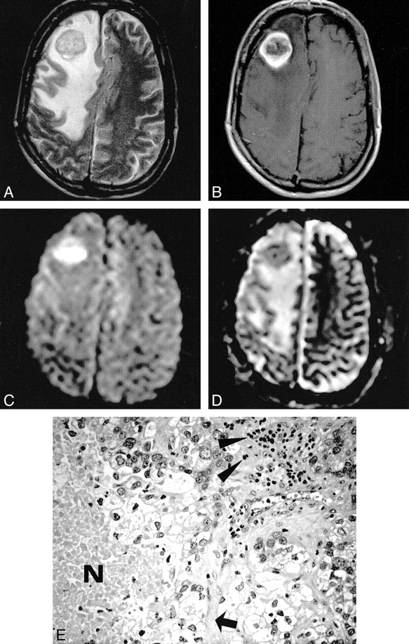
Restricted Diffusion Within Ring Enhancement Is Not Pathognomonic For Brain Abscess American Journal Of Neuroradiology

Pin On Radiography

White Matter Diseases Human Anatomy And Physiology White Matter Radiology

Leading Edge Enhancement Adrenoleukodystrophy Radiology Radiography Science Biology

Virtualmedstudent Com Tuberculosis Meningitis Radiology Radiology Imaging Meningitis

Huntington Disease The Normal Mean Fh Cc Ratio Range Is 2 2 To 2 6 As The Caudate Heads Reduce In Volume The Cc Distanc Huntington Disease Mri Brain Disease

The Use Of Magnetic Resonance Imaging In The Diagnosis And Long Term Management Of Multiple Sclerosis Neurology

Pin On Neurophotonics

Brain Tumours Imaging Brain Tumor Radiology Tumor
Posting Komentar untuk "What Does Enhancement Mean On Brain Mri"