What Cells Line The Ventricles Of The Brain
Ependymal cells ependymocytes line the brain ventricles and spinal cord canal in a continuous sheet of epithelium known as the ependyma. 3 ependymal cells called tanycytes line the floor of the third ventricle in the brain.

What Type Of Cells Line The Ventricles Of The Brain Study Com
The subventricular zone SVZ is a region situated on the outside wall of each lateral ventricle of the vertebrate brain.

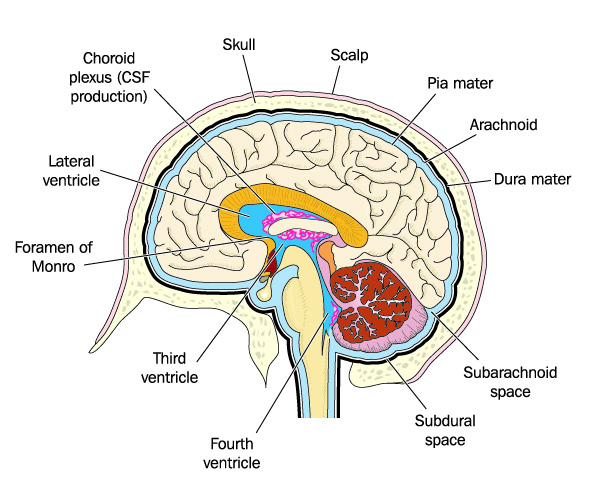
What cells line the ventricles of the brain. These cells line the cerebrospinal fluid CSF filled ventricles in the brain and the central canal of the spinal cord. The signal spreads to the atrioventricular node AV node. The co ordinated beating of cilia influences the direction of flow of the cerebro spinal fluid distribution of neuro transmitters and other messengers to the neurons. It produces cerebrospinal fluid and provides a toxin barrier to the brain and other central nervous system tissue. The ventricular system contains the lateral third and fourth ventricles whose function is to produce cerebrospinal fluid. It is present in both the embryonic and adult brain.
Ependymal cell type of neuronal support cell that forms the epithelial lining of the ventricles cavities in the brain and the central canal of the spinal cordEpendymal cells also give rise to the epithelial layer that surrounds the choroid plexus a network of blood vessels located in the walls of the lateral ventricles the two largest ventricles which occur as a pair in the cerebral. They also prune synapses. Signals carried from the AV node slightly delayed through bundle of His fibers and Purkinjie. These are nervous tissue cells with a ciliated simple columnar shape similar to some mucosal epithelial cells. The ventricles produce and contain cerebrospinal fluid CSF and the entire surface of the ventricular system is lined by an epithelial layer called the ependyma. They line cavities of brain and spinal cord and maintain cerebrospinal fluid CSF flow.
Modified muscle cells contract sending a signal to other muscle cells in the heart to contract. Ependymal cells are extremely small and line up tightly together to form. The primary neural stem cells of the brain and spinal. Learn where CSF is found. During development newly formed neurons proliferate adjacent to the canal to form a mantle layer which becomes the gray matter in the central region of. Snyder in Principles of Regenerative Medicine 2008 Definition.
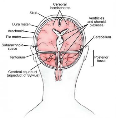
The choroid plexus and the cerebrospinal fluid that it produces are necessary. The choroid plexus is a network of capillaries and specialized ependymal cells found in the cerebral ventricles of the brain. This International journal Journal of Clinical Neuroscience publishes articles on clinical neurosurgery and neurology and the related neurosciences such as neuro-pathology neuro-radiology neuro-ophthalmology and neuro-physiology. Ependymal cells also line the ventricles of the brain that are continuous with the central canal of the spinal cord. They are involved in the production of cerebrospinal fluid which serves as a cushion for the brain moves the fluid between the spinal cord and the brain and is a component for the choroid plexus. In the ventricles these cells have small hairlike structures on them called cilia which help encourage the flow of cerebrospinal fluid.
Microglia are the brains immune cells protecting it from invaders and cleaning up debris. Ependymal cells are another glial subtype that line the ventricles of the CNS forming a permeable barrier between the cerebrospinal fluid CSF and underlying cells and also aid in the circulation of CSF through cilial beat. These types of cells are column shaped and usually line up together to form a membrane called the ependyma. Learn the ventricles of the brain along with their definition function location anatomy and cerebrospinal fluid CSF flow using labeled diagrams. But it is not the volume of growth alone that. The location of stem cells in the adult brain was later identified to be within the striatum 9 and researchers began to show that cells isolated from this region and the dorsolateral region of the lateral ventricle of the adult brain were capable of differentiating into both neurons and glia.
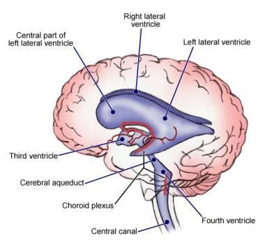
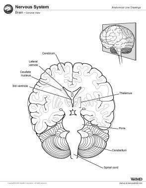
Ependymal cells line fluid-filled ventricles of the brain and the central canal of the spinal cord. The ventricles are filled with cerebrospinal fluid CSF which bathes and cushions the brain and spinal cord within their bony confines. The ependyma is composed of ependymal cells known as ependymocytes which is a type of glial cell. 2 the ependymal cells have cilia facing the cavity of the ventricles. Neural Stem Cells NSCs are the most primordial and uncommitted cells of the nervous system and are believed to give rise to the vast array of more specialized cells of the CNS and peripheral nervous system PNS. Ependymocytes line the ventricles and central canal of the spinal cord tanycytes line the floor of the third ventricle over the hypothalamic median eminence and choroidal epithelial cells cover the surface of choroidal plexus.
Grades K-6 Brain It is a card game similar to Go Fish How to play. These cells primarily produce cerebrospinal fluid CSF. The ependyma is a thin membrane lining the spinal cord and ventricles of the brain. Ependymal cells line the ventricles and secrete cerebrospinal fluid CSF. The choroid plexus serves two roles for the body. Ependymal cells line fluid-filled ventricles of the brain and the central canal of the spinal cord.
The making of the human brain from the tip of a 3 millimeter neural tube is a marvel of biological engineering. They are involved in the production of cerebrospinal fluid which serves as a cushion for the brain moves the fluid between the spinal cord and the brain and is a component for the choroid plexus. CSF is produced by modified ependymal cells of the choroid plexus found in all components of the ventricular system except for the cerebral aqueduct and the posterior and anterior horns of the lateral ventriclesCSF flows from the. Oligodendroglia cells create a fatty substance called myelin that insulates axons allowing electrical messages to travel faster. To be considered a neural stem cell in contrast to a progenitor cell ie. They also create cerebrospinal fluid and are involved in the BBB.
These findings and several others looking at cell regeneration in other parts of the brain opened up a whole new line of research about adult neurogenesis the process of the birth of neurons from neural stem cells in a mature brain. To arrive at the more than 100 billion neurons that are the normal complement of a newborn baby the brain must grow at the rate of about 250000 nerve cells per minute on average throughout the course of pregnancy. Also there is a vascular pia mater in the roofs of the third and fourth ventricles and in the medial wall of the lateral ventricle along the line of the choroid fissure. The aim of the study was to characterize cultured brain endothelial cells pericytes and glial cells from wild-type and ApoB-100 transgenic mice and to study the effect of oxidized low-density lipoprotein oxLDL on these cells. Ependymal cells are primarily known for making up a membrane called the ependyma which is a thin membrane lining the central canal of the spinal cord and the ventricles passageways of the brain. Print out ONE copy of these Brain Cards or two copies of these Brain CardsThe first set of cards is a traditional set of 52 playing cards with pictures of different brains.
In embryonic life the SVZ refers to a secondary proliferative zone containing neural progenitor cells which divide to produce neurons in the process of neurogenesis. Depending on where they are located ependymal cells also help to distribute neurotransmitters and hormones associated with the central nervous system. The journal has a broad International perspective and emphasises the advances occurring in Asia the Pacific Rim region Europe.
Choroid Plexus The Definitive Guide Biology Dictionary
Ventricles Of The Brain Overview Gross Anatomy Microscopic Anatomy

Ventricles Of The Brain Labeled Anatomy Function Csf Flow Definition Ezmed
Ventricles Of The Brain Overview Gross Anatomy Microscopic Anatomy
Ependymal Cell Anatomy Britannica
![]()
Third Ventricle Anatomy Kenhub

Brain Ventricles
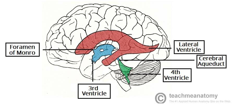
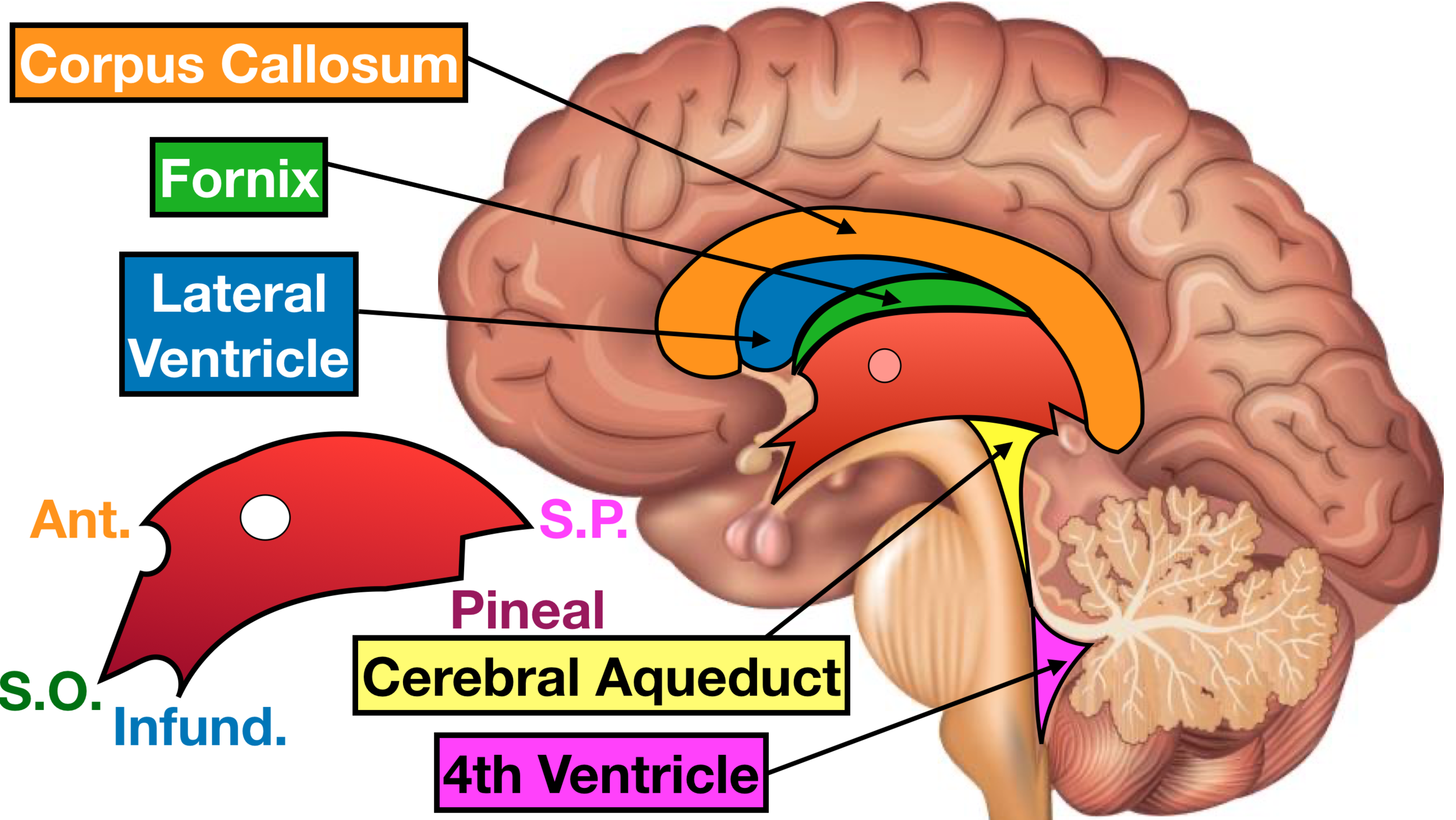
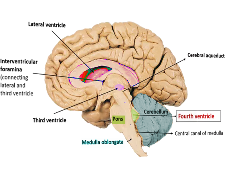
The Ventricles Of The Brain Lateral Third Fourth Teachmeanatomy
Ventricles Of The Brain Labeled Anatomy Function Csf Flow Definition Ezmed
Ventricles Of The Brain Labeled Anatomy Function Csf Flow Definition Ezmed
Neuroanatomy Fourth Ventricle Article
Ventricles Of The Brain Overview Gross Anatomy Microscopic Anatomy
Ventricles Of The Brain Overview Gross Anatomy Microscopic Anatomy
How Many Ventricles Are Present In The Human Brain Quora
Posting Komentar untuk "What Cells Line The Ventricles Of The Brain"