Hemorrhagic Rathke's Cleft Cyst
Only a few cases of apoplexy associated with RCCs have been reported and their clinical imaging surgical and pathological features are poorly understood. The clinical manifestations of RCC are characterized by headache hypopituitarism visual disorder and diabetes insipidus.
Hemorrhagic Pituitary Adenoma Versus Rathke Cleft Cyst A Frequent Dilemma Ajnr Blog

Very few articles have been published describing the details of RCC apoplexy.

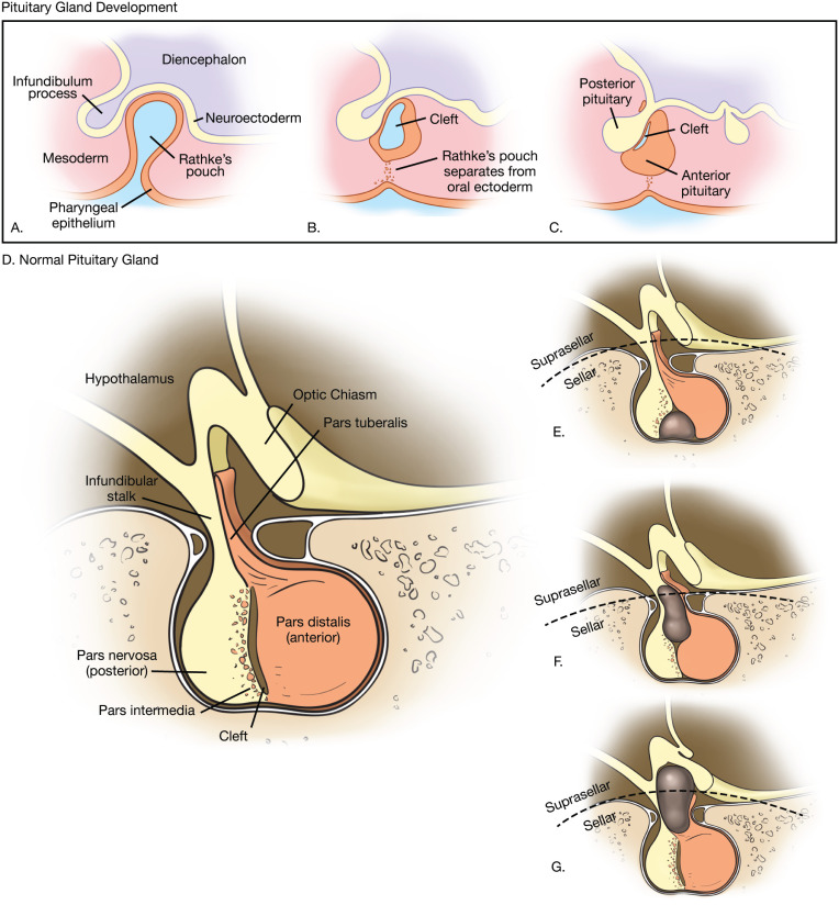
Hemorrhagic rathke's cleft cyst. Rarely RCCs hemorrhage and mimic pituitary apoplexy on presentation. Hemorrhagic and nonhemorrhagic Rathke cleft cysts mimicking pituitary apoplexy. Rathke cleft cysts are cystic sellar and suprasellar lesions arising from remnants of the embryonic Rathke pouch a structure of ectodermal origin that folds superiorly from the pharynx during the fourth week of gestation. Symptomatic Rathke cleft cysts RCCs are rare sellar and suprasellar lesions and apoplexy is one of the most unusual presentations. Rathke cleft cyst RCC is a benign epithelial cyst believed to originate from the remnants of the Rathke pouch. Rathkes cleft cysts that cause symptoms are relatively uncommon lesions accounting for less than one percent of all primary masses within the brain.
40 are purely intrasellar and 60 have suprasellar extension. Rathke cleft cysts RCCs are infrequently symptomatic and apoplexy is one of the most unusual presentations. Rathkes cleft cysts RCCs are benign sellar andor suprasellar lesions 1 originating from the remnants of Rathkes pouch. 3 Luschka in 1860 and then Goldzieher in 1913 published the first cases of RCCs as. Purely suprasellar location although reported is rare. A Rathke cleft cyst develops from a piece of the fetus developing Rathke pouch which ultimately becomes part of the pituitary gland.
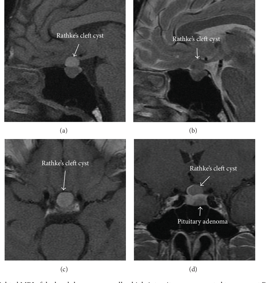
Hemorrhagic Rathkes cleft cyst In this case craniopharyngioma was felt to be less likely since a high percentage of these calcify and typically display thick-walled solid or nodular enhancement. 9 Typical imaging findings include a nonenhancing noncalcified intrasellarsuprasellar cyst with an intracystic nodule. Rathkes cleft cyst is found in 1233 of routine autopsies 1 2. Rathke cleft cysts RCCs are infrequently symptomatic and apoplexy is one of the most unusual presentations. On imaging a Rathkes cleft cyst is seen as a well defined non enhancing midline cyst within the sella arising between the anterior and intermediate lobes of the pituitary. Rathkes cleft cysts sometimes occur together with pituitary adenomas.
2 They are named after the German anatomist Martin Heinrich Rathke who described the evagination process of the anterior foregut in 1838. Only a few cases of. In the cases that have been reported intracystic hemorrhage has been a consistent finding. In daily practice this situation may be particularly confusing in a young woman with mild hyperprolactinemia whose symptoms are frequently hidden by taking contraceptive pills. Imaging examinations usually indicate a lesion located in the sellar or suprasellar region. Rathke cleft cysts are non-cancerous fluid-filled growths that develop between the parts of the pituitary gland at the base of the brain.
Developed by renowned radiologists in each specialty STATdx provides comprehensive decision support you can rely on - Rathke Cleft Cyst. They can occur at any age although most are identified in adults. Imaging revealed a 12 cm cystic pituitary mass consistent with a hemorrhagic Rathkes cleft cyst. Only a few cases of apoplexy associated with RCCs have been reported and their clinical imaging surgical and pathological features are poorly understood. We present a 49-year-old male patient who presented with a 3-month history of progressive frontotemporal headaches. They develop while a fetus is growing in the womb.
Rathke cleft cyst RCC is an epithelium-lined benign congenital cystic lesion found in the sellar or suprasellar area. Macroadenoma was felt to be less likely due to lack of cavernous sinus invasion and the uniformly thin wall of this lesion. 12 rows Rathke cleft cysts are remnants of the Rathke pouch a structure of ectodermal origin formed. In the cases that. Occasionally this remnant gives rise to a large cyst called the Rathkes cleft cyst RCC. Their incidence has been underestimated before.
It is concluded that MRI is an efficient tool for diagnosis allowing appropriate medical decision making in Rathkes cleft cyst cases based on a retrospective review of 12 cases and a review of the literature. In the sella Rathke pouch gives rise to the adenohypophysis anteriorly and intermediate lobe of the pituitary gland posteriorly. Rathke cleft cyst RCC apoplexy is an uncommon type of lesion that is challenging to diagnose without histopathological samples. Differentiation with MR imaging of a cystic or hemorrhagic pituitary adenoma from a Rathke cleft cyst RCC remains a common issue. Rathke cleft cysts RCCs are benign sellar and suprasellar lesions commonly presenting as asymptomatic incidental findings. To report a case of uncommon presentation of hemorrhagic Rathkes cleft cyst RCC extending into the cavernous sinus causing diplopia.
9 12 Depending on its cystic content and the presence of an associated intracystic nodule an RCC may show various signal. The correlation of gross and pathologically identified hemorrhagic products in the patients included provides a very good correlation with the radiographic findings and further helps clarify the fact that although most Rathkes cleft cysts have evidence of high proteinaceous fluid which can mimic hemorrhage there are certain cysts that present with an actual hemorrhage. Up to 10 cash back Malignant transformation of a Rathkes cleft cyst has never been described. PURPOSE Rathkes cleft cysts are non neoplastic lesions of the sellar area that seldom are symptomatic. To review a series of hemorrhagic RCCs for physicians encountering this rare presentation.

Rathke S Cleft And Other Pituitary Cysts Pituitary World News
T2 Hypointense Signal Of Rathke Cleft Cyst American Journal Of Neuroradiology

2
Fig 1 Mr Imaging Findings Of Rathke S Cleft Cysts Significance Of Intracystic Nodules American Journal Of Neuroradiology


Rathke Cleft Cyst Neupsy Key
Fig 1 Hemorrhagic Pituitary Adenoma Versus Rathke Cleft Cyst A Frequent Dilemma American Journal Of Neuroradiology

Pdf Gh Producing Pituitary Adenoma And Concomitant Rathke S Cleft Cyst A Case Report And Short Review Semantic Scholar

Scielo Brasil Bilateral Acute Visual Loss From Rathke S Cleft Cyst Apoplexy In A Patient With Dengue Fever Bilateral Acute Visual Loss From Rathke S Cleft Cyst Apoplexy In A Patient With
Rathke Cleft Cyst Presenting As Recurrent Pituitary Apoplexy An Unusual Case Presentation And Clinical Course
Rathke Cleft Cyst Radiology Key

Mris Of Rathke S Cleft Cysts A Coronal I And Sagittal Ii Download Scientific Diagram
Differentiation Between Cystic Pituitary Adenomas And Rathke Cleft Cysts A Diagnostic Model Using Mri American Journal Of Neuroradiology


Rathke S Cleft Cyst Operative Neurosurgery
Fig 6 Differentiation Between Cystic Pituitary Adenomas And Rathke Cleft Cysts A Diagnostic Model Using Mri American Journal Of Neuroradiology

Posting Komentar untuk "Hemorrhagic Rathke's Cleft Cyst"