Pet Brain Imaging Metabolic Evaluation
An ischemic stroke is the most common type. 78608 Brain imaging PET metabolic evaluation 78609 Brain imaging PET perfusion evaluation HCPCS Code Description None None Table 3.

Tau And Amyloid Positron Emission Tomography Pet Scans In A Typical Download Scientific Diagram
A right brain stroke happens when blood supply to the right side of the brain is stopped.
Pet brain imaging metabolic evaluation. Physicians who specialize in the performance and interpretation of neuroimaging in the clinical setting are neuroradiologists. The most common use of PET is in the detection of. The PET imaging tracers help in differentiating dementia syndromes which do not have overlap of the underlying pathological process. Chest Imaging CPT HCPCS and Diagnoses Codes You may access the Chest Imaging Diagnoses Codes 932 here. PET measures emissions from radioactively labeled metabolically active chemicals that have been injected into the bloodstream. Neuroimaging or brain imaging is the use of various techniques to either directly or indirectly image the structure function or pharmacology of the nervous systemIt is a relatively new discipline within medicine neuroscience and psychology.
Laboratory Evaluation of Dementia Test Intended diagnosis Use Comments Psychometric testing All dementias especially MCI FTD In appropriate clinical context Virtually required for MCI mild AD and FTD. 78608 Brain imaging positron emission tomography PET. Generally before these tests are ordered a physical examination is done to determine whether there are neurological changes that suggest the presence of a brain tumor. A positron emission tomography PET scan is an imaging test that helps reveal how your tissues and organs are functioning. When performing SPM analysis for different patient. Binder et al 1999.
The following codes may be applicable to chest imaging and may not be all-inclusive. Ischemic and hemorrhagic. It is now almost 10 years since brain functional imaging studies first suggested that cerebral blood flow and metabolism may vary across different cortical regions during the conscious resting state being somewhat greater in the medial parietal medial occipital and mid-dorsolateral prefrontal areas Gur et al 1995. Positron emitting tracer is injected into the body which emits positrons causing annihilation that results two gamma rays. Disparities in Statin Use During Outpatient Visits of Adults Age 18 to 64 Years With Coronary Heart Disease in the Medicaid Population in the United States from the National Ambulatory Medical Care Survey Database 2006 to 2015. Then these signal is transfer to amplifier and other.
Performs fusion to a CT andor MR dataset. A PET scan uses a radioactive drug tracer to show this activity. Limited area eg chest headneck. These rays are detected by opposing detectors. It also does some thought processing help us know body position and judge space and distance. The scans range in time from 20-40 minutes but you will be asked to arrive early to allow the tracer time to adhere to areas of high metabolic activity.
Paralysis of a leg often results from damage to the peripheral spinal nerves. Statistical Parametric Mapping SPM is a computational approach for analysing functional brain images like Positron Emission Tomography PET. According to a 2020 study people with high levels of. 78811 Tumor imaging positron emission tomography PET. They reveal the brains anatomy including the integrity of brain structures and their interconnections. Combined with FlowMotion it is designed to.
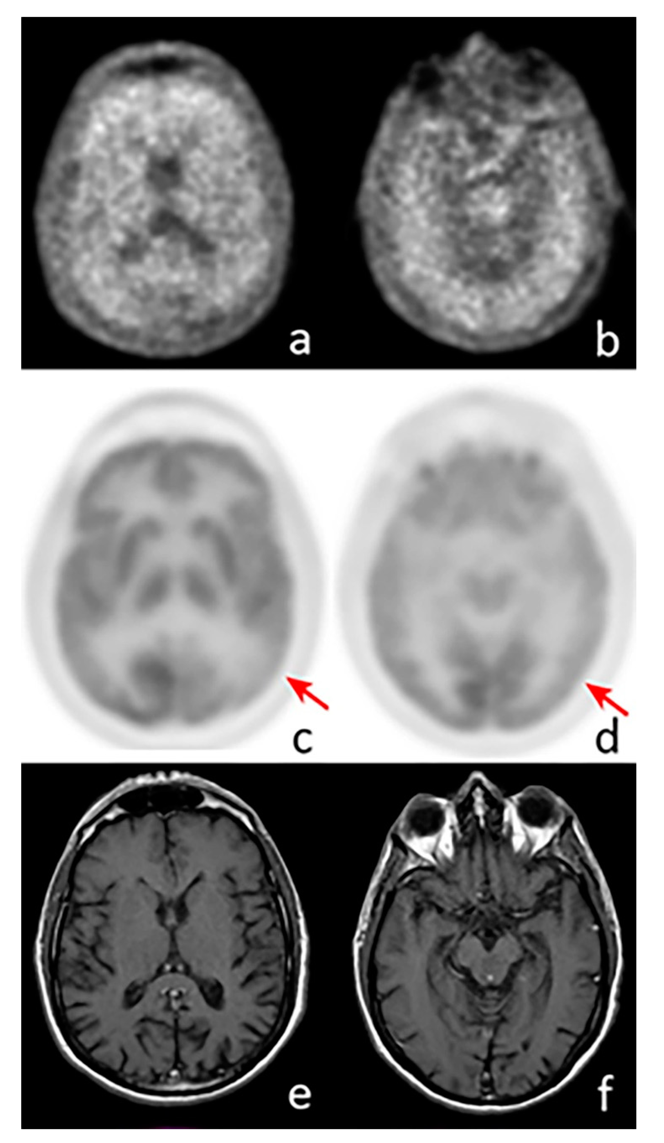
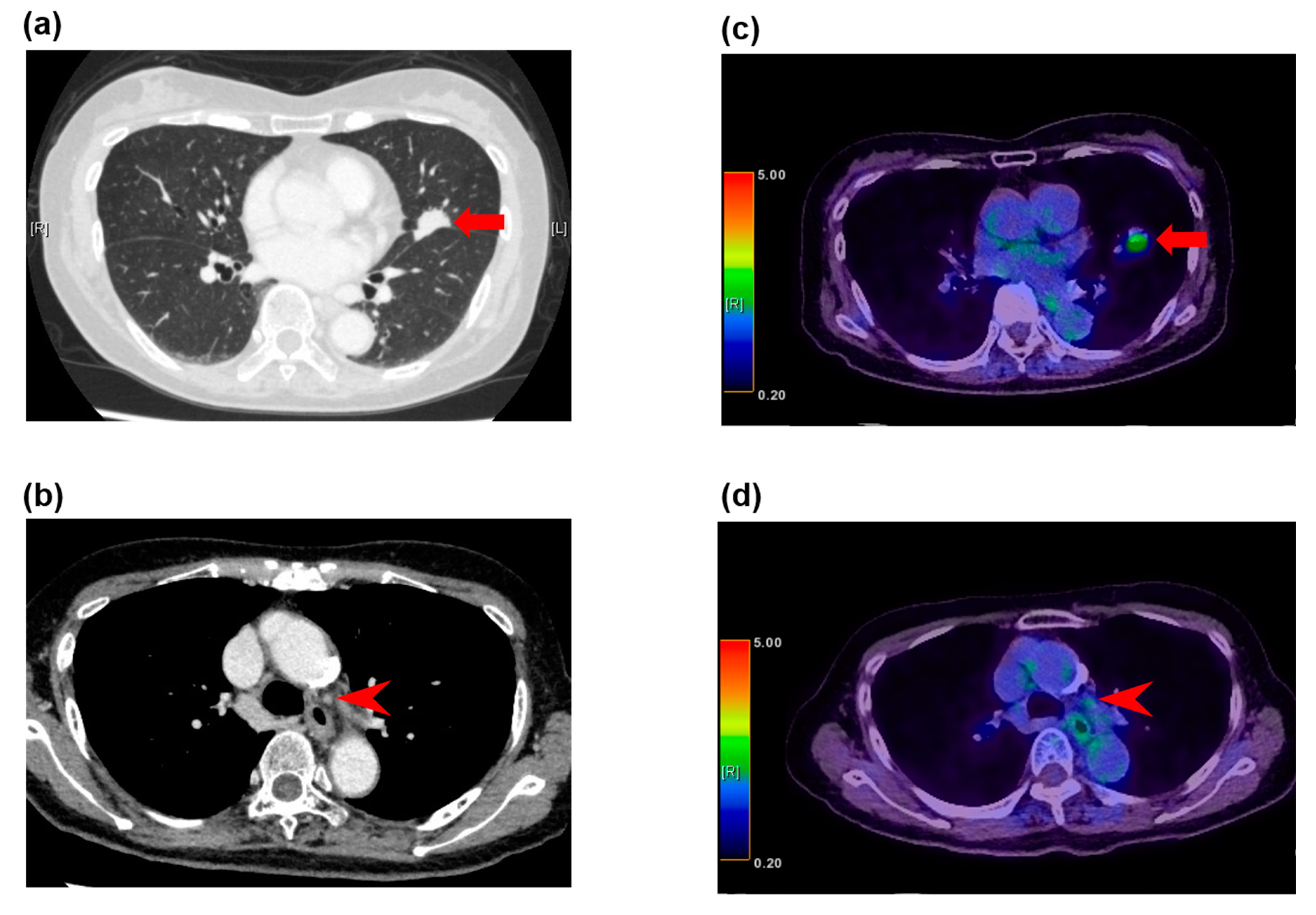
Current neuroimaging techniques reveal both form and function. Brain imaging can also connect certain mental health issues to biological causes as well. Positron emission tomography PET is being increasingly used for diagnosis staging and follow-up of various malignancies. The amyloid deposition in the brain can be detected years before the onset of clinical symptoms. FDG-PET of the brain demonstrates a hypermetabolic focus within the right medial temporal lobe lesion B. PET of the body demonstrates a hypermetabolic focus in the left lung E consistent with biopsy-proved small-cell lung cancer.
The newest tools show how different regions of the brain connect and communicate. For example syngo PET Database Comparison. In general PET scans may be used to evaluate organs andor tissues for the presence of disease or other conditions. Brain positron emission tomography is a form of positron emission tomography PET that is used to measure brain metabolism and the distribution of exogenous radiolabeled chemical agents throughout the brain. Biograph Vision can help you optimize your clinical operations with quality images and efficient workflow. 13 The purpose of this article was to review the role of brain PET imaging in the diagnosis of AD.
Depending on the type of procedure performed the system can be configured so syngovia. In neurology PET brain imaging is becoming more utilized due to the aging population and development of new biomarkers. This study is a prospective evaluation of regional and whole-body PET imaging for staging lung cancer carried out by the Northern California PET Imaging Center California in 99 patients. WHAT IS PET PET is Nuclear medicine functional imaging technique for metabolic processes functions in the human body. It has been studied in the evaluation of various tumors including but not limited to solitary pulmonary nodules nonsmall cell lung carcinoma lymphoma melanoma breast cancer and colorectal cancer 1 7. There are two main types of stroke.
PET may also be used to evaluate the function of organs such as the heart or brain. 78492 Myocardial imaging positron emission tomography PET perfusion multiple studies at rest andor stress. A brain tumor can be diagnosed using imaging tests that view the structure of the brain along with a biopsy which can carefully assess a sample of a suspected brain tumor under a microscope. Localized proton MR spectroscopy MRS of the human brain first reported more than 20 years ago13 is a mature methodology that is used clinically in many medical centers worldwide for the evaluation of brain tumors While there have been studies of human brain tumors using heteronuclei such as phosphorus 31 P and sodium 11 Na56 by far the most. Baltzers aim to further improve upon the quality speed and transparency of publishing in the European Journal of Radiology. Paralysis of a front leg is usually associated with injury to the nerve roots in the neck or shoulder injury to the network of nerves found deep in the armpit brachial plexus or injury to the radial median musculocutaneous or ulnar nerve in the leg.
The patient was in remission following treatment with intravenous immunoglobulin infusions oral steroids and. They elucidate its chemistry physiology and electrical and metabolic activity. He has published 200 papers book chapters and reviews in the field of imaging and he and his co-workers have repeatedly received national and international awards. May be essential if medico-legal complications are possible CBC electrolyte panel calcium SUN creatinine glucose Common metabolic. With the markets highest effective sensitivity at 100 cpskBq 3 and the fastest time of flight in the industry 4 Biograph Vision can not only reduce scan time and injected dose to boost productivity it can also improve image quality. Starts the appropriate reading environment.
The right side of the brain is in charge of the left side of the body.

18f Florbetapir Pet Mri For Quantitatively Monitoring Myelin Loss And Recovery In Patients With Multiple Sclerosis A Longitudinal Study Eclinicalmedicine

Brain Mri Brain Fdg Pet Ct And Hypometabolic 3d Ssp Maps For 3 Download Scientific Diagram

Effect Of Deep Brain Stimulation On The Brain Metabolic Pattern In Patients With Depression Comparison Of Pre And Post Therapy Pet Ct Scans A Pilot Study Journal Of Nuclear Medicine

Molecular Imaging Of Brain Tumors With Pet Typical Pet Tracers Used Download Scientific Diagram

Sequential Fdg Pet And Mri Findings In A Case Of Smart Syndrome Seizure European Journal Of Epilepsy
Diagnostics Free Full Text The Additional Value Of 18f Fdg Pet And Mri In Patients With Glioma A Review Of The Literature From 2015 To 2020 Html

References In Pet Mri Multiparametric Imaging Of Brain Tumors European Journal Of Radiology
Ijms Free Full Text The Added Value Of Diagnostic And Theranostic Pet Imaging For The Treatment Of Cns Tumors Html
Cancers Free Full Text Use Of Pet Imaging In Neuro Oncological Surgery Html
Ijms Free Full Text Positron Emission Tomography Pet And Neuroimaging In The Personalized Approach To Neurodegenerative Causes Of Dementia Html

F18 Fet Pet Mri Scan Of 60 Year Old Male Patient With Glioblastoma Download Scientific Diagram
Diagnostics Free Full Text 18f Fdg Pet Imaging Patterns In Autoimmune Encephalitis Impact Of Image Analysis On The Results Html
Diagnostics Free Full Text Recent And Current Advances In Fdg Pet Imaging Within The Field Of Clinical Oncology In Nsclc A Review Of The Literature Html
Detection Of Clinically Silent Brain Lesions In 18f Fdg Pet Ct Study In Oncological Patients Analysis Of Over 10 000 Studies Scientific Reports
Posting Komentar untuk "Pet Brain Imaging Metabolic Evaluation"